Stem cell-derived cardiomyocytes come to IEMR

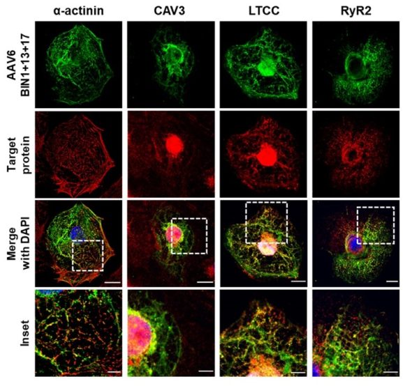
T-tubules grown in iPSC-derived cardiomyocytes align with the sarcomeric protein a-actinin, as well as the t-tubular proteins caveolin-3 (CAV3), the L-type Ca2+ channel (LTCC), and the nearby Ryanodine Receptor (RyR). From Perdreau-Dahl et al., Circ Res, 2023.

IEMR-researchers have acquired the know-how and infrastructure to generate mature iPSC-derived cardiomyocytes.

The heartbeat is triggered by the coordinated contraction and relaxation of cardiomyocytes. These cells are formed during the heart’s embryonic development and are initially highly proliferative. However, in the adult heart, cardiomyocytes do not divide, and this prevents their long-term culture as a cell line. This has traditionally limited our ability to investigate adult cardiomyocyte physiology and pathophysiology, particularly for human cardiomyocytes which are not widely available.

However, new techniques are overcoming these limitations. Stem cell research has witnessed significant developments over the last two decades, and now allows the reprogramming of human somatic cells into induced pluripotent stem cells (iPSCs). These iPSCs can be further re-differentiated into cardiomyocytes. Human iPSC-derived cardiomyocytes have already proven to be an excellent resource for understanding cardiac biology. In addition, human iPSC cardiomyocytes can be used to investigate the mechanisms of genetic diseases by examining cardiomyocytes derived from affected patients. With recent developments in gene editing technology, iPSCs can be employed to model or correct genetic diseases, and these cells have great potential for use in cell-based therapies that restore damaged heart tissues. Moreover, human iPSC-derived cardiomyocytes are also now regularly used in drug testing for evaluation of cardiac safety.
With recent developments in gene editing technology, iPSCs can be employed to model or correct genetic diseases, and these cells have great potential for use in cell-based therapies that restore damaged heart tissues.
-

At IEMR, recent work in the group of Professor Bill Louch has been employing human iPSC-derived cardiomyocytes in a number of settings. Postdoc Harmonie Perdreau-Dahl and Researcher Pugal Erusappan have used these cells to examine the mechanisms of t-tubule development (see figure). Postdoc Jia Li and PhD student Magnhild Sekse-Erdal have meanwhile been employing iPSC-derived cardiomyocytes to investigate the function of individual contractile units, called sarcomeres, to understand how mutations in the elastic protein titin alter sarcomere function in patients.

Until the past year, collaborating groups have provided the iPSC-derived cardiomyocytes that IEMR scientists have been using in their experiments. This has now changed, as Drs. Li and Erusappan have acquired the know-how and infrastructure to prepare our own cells. While it is challenging to generate mature iPSC-derived cardiomyocytes that resemble those in the adult human heart, considerable progress has already been made. Indeed, Li and Erusappan have observed the presence of well-formed sarcomeres and t-tubules in these cells.

We look forward to bringing iPSC-derived cardiomyocytes into an expanding project portfolio at IEMR, as we aim to increase the translational nature of our science and develop new human therapies.