Markus Borge Harbo; Einar Sjaastad Nordén; Jagat Narula; Ivar Sjaastad and Emil Knut Stenersen Espe
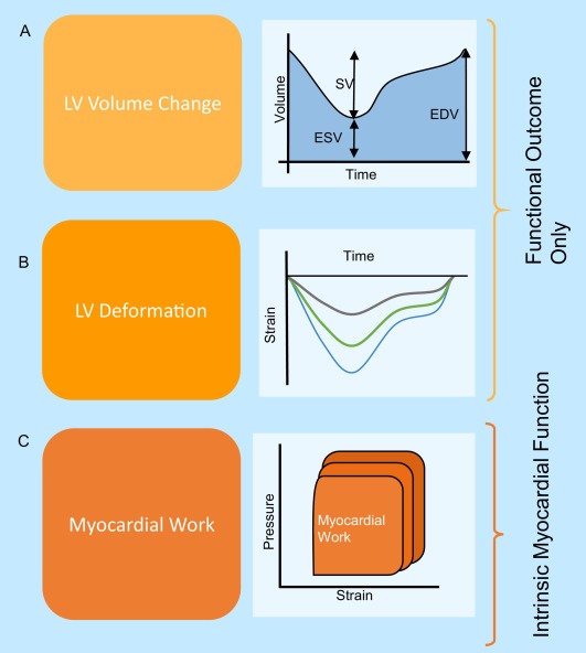
In heart failure (HF) management, noninvasive quantification of left ventricular (LV) function is rapidly evolving. Deformation parameters, such as strain, continue to challenge the central role of ejection fraction (EF) in diagnosis and prognostication of LV dysfunction in HF. The increasing recognition and use of deformation parameters motivates a conceptual discussion about what makes a parameter clinically valuable. To do this, we introduce a framework for parameter evaluation. The framework considers three aspects that are important for parameter value; 1) how these parameters couple with underlying myocardial function; 2) the evidence base of the parameters; and 3) the technical feasibility of their measurement. In particular, we emphasize that the coupling of each parameter to the underlying myocardial function (aspect 1) is crucial for parameter value. While EF offers information about cardiac dysfunction trough measuring changes in LV volume, deformation parameters more closely reflect underlying myocardial processes that contribute to cardiac pumping function. This is a fundamental advantage of deformation parameters that could explain why a growing number of studies supports their use. A close coupling to underlying function is, however, not sufficient for high clinical value by itself. A parameter also needs a strong evidence base (aspect 2) and a high degree of technical feasibility (aspect 3). By considering these three aspects, this review discusses the present and potential clinical value of EF and deformation parameters in HF management.
Read the article in Progress in Cardiovascular Diseases.
Volume 63, Issue 5, September–October 2020, Pages 552-560

Figure: Measuring myocardial function on different levels. A) Volume changes is the net outcome of LV function. B) LV deformation gives a detailed reflection of left ventricular function, for instance parametrized by strain. C) Myocardial work is estimated from the area spanned by a pressure-strain loop. The area of the pressure-strain loop acknowledges changes in load. LV = left ventricle, SV = stroke volume, ESV = end systolic volume, EDV = end diastolic volume.

