Preclinical MR Core Facility

Our preclinical MR core facility offers a variety of advanced imaging methods, with a particular focus on the assessement of cardiac structure and function.


... non-invasive imaging capabilities, with superior spatial and temporal resolution
Preclinical MR Core Facility, IEMR

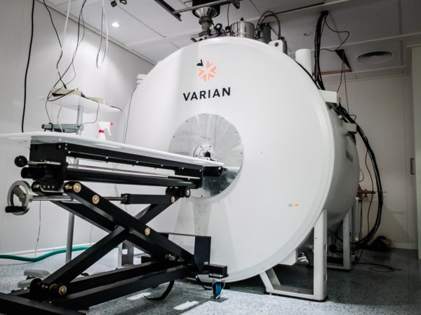
MR scanner at our core facility consists of a 9.4 T magnet from Varian/Agilent interfaced to the most updated spectrometer Avance Neo from Bruker that allows unparalleled flexibility for MRI scanning of rodents up to 800 g and other samples.

The system includes full set of RF coils optimized for cardiac, neuro- imaging or full body imaging. There are also coils to investigate metabolism via 31P imaging.

Our MR scanner offers unique non-invasive imaging capabilities, with superior spatial and temporal resolution imaging which is not achievable with existing other instruments.

Booking information

Cardiac Imaging |Ex-Vivo Imaging |
Metabolism | Neuro Imaging | Prostate Imaging

Cardiac Imaging

CINE imaging with Compressed Sensing (CS)

Cine cardiac imaging is the primary technique by which cardiovascular magnetic resonance (CMR) imaging characterizes regional and global contractile function of the heart and resulting blood flow through the great vessels. It has evolved to be the gold standard in both clinical and preclinical cases to assess heart structure and function.

The standard method to acquire Cine is slice by slice in short axis view, but it is time consuming. Our lab has developed fast imaging technique, which speeds up to 8 x. (McGinley et al., 2019)

Figure: Compressed sensing for speed acquisition.

Figure: 4D cine and LGE

Late gadolinium enhancement (LGE)

Late gadolinium enhancement is a technique based on differences in the volume of distribution of gadolinium, an extracellular agent. It is widely used in cardiac magnetic resonance imaging for cardiac tissue characterization, in particular, the assessment of regional scar formation and myocardial fibrosis.  The presence of scar and/or fibrosis in the myocardium showed hyper intensity in LGE images.

Our facility has developed a fast protocol, which is capable to acquire full heart high quality LGE in 3 minutes. It’s also feasible to acquire CINE and LGE at one session in about 10 minutes.


Figure: LGE of infarct rat at one day after operation

Flow and Tissue Phase Mapping

Phase-contrast MRI is a versatile MRI technique, which routinely is used to measure blood flow (flow-encoded MRI) or myocardial function (tissue phase mapping). Our core facility has developed 3D flow phase-contrast MRI (Skårdal, Espe, Zhang, Aronsen, & Sjaastad, 2016).

We also established phase-contrast MRI as a robust tool for measuring the regional function of the myocardium as well, in particular strain, stress and work (Emil K S Espe et al., 2015; Emil K S Espe et al., 2017; Emil Knut Stenersen Espe, Aronsen, Norden, Zhang, & Sjaastad, 2019)

Figure: Strain and Stress in normal heart (left) and infarcted heart (right)
Left: Regional work, infarct without heart failure (left) and without heart failure (right). Right: Flow and Tissue Phase Mapping.

Figure: Native T1 map of an infarct rat.

T1 mapping

Increased fibrotic growth in extracellular matrix is associated with stiffening of the ventricle, leading to diastolic and systolic, dysfunction. Detecting myocardial fibrosis is of importance for risk stratification, contributes to understanding the mechanisms responsible for fibrosis initiation, progression.

T1 mapping is a technique to quantify the exact T1 of the tissue. Post contrast T1 mapping allows the detection of variations in gadolinium distribution within the tissue with higher resolution than replacement fibrosis imaging, i.e. LGE. Combining precontrast/native T1, postcontrast T1 and hematocrit, the extra cellular volume (ECV) fraction can be calculated.

Figure: Post-contrast T1 map (left) and ECV map (right) of the same rat.

Diffusion Tensor Imaging (DTI)

DTI allows us to quantify the degree and direction of water diffusion in tissue. The technique exploits diffusion encoding gradients in several directions so as to determine the degree of diffusion in 3D. This can be used to determine tissue properties such as myofiber architecture and extra-cellular volume (ECV). This is currently being performed ex-vivo, however the sequence is being adapted for future in-vivo use.

The cardiac muscle fiber structure of the healthy rat heart, measured ex vivo using Diffusion Tensor Magnetic Resonance Imaging (Bård Andre Bendiksen, IEMR)

Magnetic Resonance Elastography (MRE)

Magnetic Resonance Elastography (MRE) is a rapidly developing technology for quantitatively assessing the mechanical properties of tissue.

Currently, MRE is used to detect stiffening of the liver caused by fibrosis and inflammation in chronic liver disease. But MRE is also being evaluated as a noninvasive way to diagnose diseases in the heart.

Our researcher is developing in vivo MRE both can be used both in clinical and preclinical setup. We are closely collaborate with Mayo Clinic and The University of Illinois at Chicago.

Figure: Stiffness map of gel phantom.

Ex-Vivo Imaging

With our special designed RF coil, we can retrieve super-high resolution 10-20 µm images within reasonable time.

It renders anatomy in great detail. The following examples are surgical anatomy of the superior orbit (Krohn-Hansen et al., 2015) and the spinal cord from mini-pig (Züchner et al., 2019).

Animation: High resolution of ex-vivo heart.


Left: Anatomy of the superior orbit of the eye, resolution 20 x 20 x 20 um- Right: Damaged spinal cord of a mini pig

Metabolism

Liver fat and body fat both play a vital role of reflecting body health. It’s closely related to body metabolism, especially lipid metabolism. By using specially tailored chemical shifting imaging technique, we can quantify the fat content in the liver. Whole body fat is also easily can be obtained by T2 weighted images (Sokolova et al., 2019).
MRS  and MRI provide the unique opportunity for in vivo assessment of several fundamental events in tissue metabolism without the use of ionizing radiation like PET imaging.  Of particular interest, phosphate metabolites that are involved in ATP generation and utilization can be quantified noninvasively by phosphorous-31 (31P) MRS/MRI.
Animation: Whole body fat quantification. 
Figure: Liver Fat Imaging: Water only image (left) and Fat only image (right)
Figure: 31P spectroscopy of a healthy rat leg

Neuro Imaging

We are also equipped with phase array neuroimaging RF coils which are designed for taking neuro imaging from mice and rats. We have studied with animal models with neuro disease varies from glioma, epilepsy and stroke (Mughal et al., 2018).

Prostate Imaging

The management of prostate cancer is challenging because the disease has variable clinical and pathologic behavior. Orthotopic mouse prostate cancer models with human cancer cells implanted in the mouse prostate or transgenic models have been used to study chemopreventive treatment efficacy. Ability to non-invasively determine the size and volume of a mouse’s prostate gland as well as tumor will help investigators in the diagnosis and evaluation of the efficacy of different treatment strategies in prostate cancer. Our 9.4 T MRI provides high-resolution images, allowing a good delineation of prostate glands and its tumor.

Contact


Senior Engineer Lili Zhang, PhD

phone: (+47) 230 16844

or (+47) 230 16849

e-mail: lili.zhang@medisin.uio.no

Location


Oslo University Hospital, Ullevål, Building 25,
Kirkeveien 166, NO-0407 Oslo, Norway

Booking information


Selected publications

Espe, E. K. S., Aronsen, J. M., Eriksen, G. S., Zhang, L., Smiseth, O. A., Edvardsen, T., . . . Eriksen, M. (2015). Assessment of regional myocardial work in rats. Circulation. Cardiovascular imaging, 8, e002695. doi:10.1161/CIRCIMAGING.114.002695
Espe, E. K. S., Aronsen, J. M., Eriksen, M., Sejersted, O. M., Zhang, L., & Sjaastad, I. (2017). Regional Dysfunction After Myocardial Infarction in Rats. Circulation. Cardiovascular imaging, 10. doi:10.1161/CIRCIMAGING.116.005997
Espe, E. K. S., Aronsen, J. M., Norden, E. S., Zhang, L., & Sjaastad, I. (2019). Regional right ventricular function in rats: a novel magnetic resonance imaging method for measurement of right ventricular strain. American Journal of Physiology. Heart and Circulatory Physiology, 318(1), 143-153. doi:http://dx.doi.org10.1152/ajpheart.00357.2019
Krohn-Hansen, D., Zhang, L., Haaskjold, E., Meling, T. R., Nicolaissen, B. r., & Sjaastad, I. (2015). Surgical anatomy of the superior orbit on ultra-high-resolution MRI at 9.4 Tesla. Journal of Plastic Surgery and Hand Surgery, 49(5), 284-288. doi:10.3109/2000656X.2015.1041969
McGinley, G., Bendiksen, B. A., Zhang, L., Aronsen, J. M., Norden, E. S., Sjaastad, I., & Espe, E. K. S. (2019). Accelerated magnetic resonance imaging tissue phase mapping of the rat myocardium using compressed sensing with iterative soft-thresholding. PLoS ONE, 14(7), e0218874. doi:10.1371/journal.pone.0218874
Mughal, A. A., Zhang, L., Fayzullin, A., Server, A., Li, Y., Wu, Y., . . . Vik-Mo, E. O. (2018). Patterns of Invasive Growth in Malignant Gliomas—The Hippocampus Emerges as an Invasion-Spared Brain Region. Neoplasia, 20(7), 643-656. doi:10.1016/j.neo.2018.04.001
Skårdal, K., Espe, E. K. S., Zhang, L., Aronsen, J. M., & Sjaastad, I. (2016). Three-Directional Evaluation of Mitral Flow in the Rat Heart by Phase-Contrast Cardiovascular Magnetic Resonance. PLoS ONE, 11(3), e0150536. doi:10.1371/journal.pone.0150536
Sokolova, M., Sjaastad, I., Louwe, M. C., Alfsnes, K., Aronsen, J. M., Zhang, L., . . . Yndestad, A. (2019). NLRP3 Inflammasome Promotes Myocardial Remodeling During Diet-Induced Obesity. Frontiers in Immunology, 10. doi:10.3389/fimmu.2019.01621
Züchner, M., Lervik, A., Kondratskaya, E., Bettembourg, V., Zhang, L., Haga, H. A., & Boulland, J.-L. (2019). Development of a Multimodal Apparatus to Generate Biomechanically Reproducible Spinal Cord Injuries in Large Animals. Frontiers in Neurology, 10(223). doi:10.3389/fneur.2019.00223